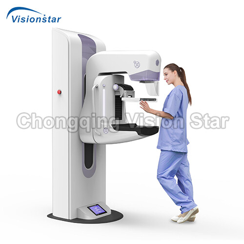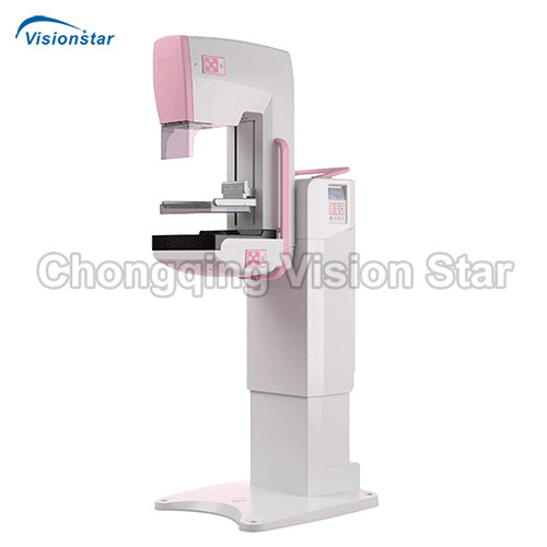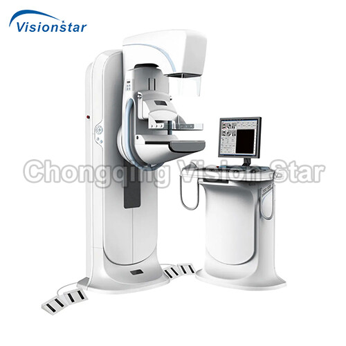
Mega 600 Digital Mammography System
$0.00
Mega 600 Digital Mammography System
Feature:
Application: It is specially used for human breast tissue photography to obtain tissue images for breast disease diagnosis and breast cancer screening. It integrates great image quality, flexible operation, good examination experience and beautiful design in one system. It is a special designed product for women healthcare and it will bring a brand new experience on digital diagnosis of breast.
* Simplify the Workflow:
One key positioning, move to the designated position automatically;
One key exposure, full-automatic AEC,
All parameters settings and filter selection are completed automatically;
Auto-mirror, body posture, position memory and mirror placement.
* Friendly Interface:
The software with friendly interface;position show by humanoid schematic diagram,
automatic generation ofstandardized examination protocol;large color LCD screen for convenience of observation and operation.
* Rich Options:
Support 1.5X and 1.8X magnification radiography component and different type options of oppression disc.
Support bar code reader and fingerprint recognition module; Let the cases input and department management into the era of intelligent.
* Open the Interface for Easy Interconnection:
It is compatible with DICOM3.0, which can seamlessly interconnect with the HIS/RIS/PACS, and automatically generated the worklist, and it can connect with remote consultation system.
* Configuration (Standard 1 PC)
- Flat Panel Detector (FDP)
- X-ray tube
- High voltage generator
- Rack structure
- Collimator
- High voltage cable
- Compression plate
- Integrated workstation
- Image acquisition software of medical mammography

 |
 |
 |


|
Technical Data |
|
|
Rack System |
|
|
Rack structure |
vertical C-arm type |
|
C-arm rotation range |
+ 195° to – 155° |
|
C-arm vertical movement range: |
630mm-1440mm |
|
C-arm can be locked at any position throughout the stroke |
|
|
The rack display screen can display the current position and shooting position information, and has the function of one key in place, which can change the preset position with one key |
|
|
Distance between focus and image receiving area (FID) |
660 mm |
|
High Voltage Generator Set (imported) |
|
|
With exposure control system, support key exposure or handbrake exposure. |
|
|
Type |
High frequency inverter type |
|
X-ray tube voltage |
20-40kV (step 0.5kV) |
|
X-ray tube current |
10mA-160mA |
|
Maximum output power |
5kW (40kV, 125mA) |
|
Nominal power |
4.8kW (30kV, 160mA, 0.1s) |
|
Radiograph time |
0.005-10s |
|
mAs |
0.5mAS~560mAs |
|
Minimum mAs |
0.5mAs (50ms 10mA) |
|
Maximun mAs |
560mAs |
|
Minimum mAs in AEC |
6.3mAs |
|
Exposure control |
Manual, semi-auto,auto |
|
Radiating mode |
Cross-ventilation |
|
X-ray tube assembly (imported) |
|
|
Double speed rotating anode |
|
|
Tube rated voltage |
40kV |
|
Anode heat capacity |
300KHU |
|
Anode rotating speed |
10000rpm |
|
Anode material |
Molybdenum |
|
Nominal focus size |
Small focus 0.1mm, large focus 0.3mm |
|
Flat panel detector component |
|
|
Imaging material |
amorphous silicon |
|
The image size |
240mm × 300mm |
|
Conventional spatial resolution |
7.0lp/mm |
|
Pixel matrix |
4096 x 3072 |
|
A/D conversion |
16 bit |
|
Pixel size |
77μm |
|
Mammography compression device |
|
|
Mammography Compression Apparatus Compression motion control is manual and electric, and the extrusion action can be released at any time during the extrusion process. In the event of a power failure, the compressor pressure plate may release pressure. |
|
|
Compression plate movement range: Under the ordinary photographic platform, the movement range is 245mm. Under the 1.5x magnification photographic platform, the range of motion is 150mm, and under the 1.8x magnification, the range of motion is 75mm. |
|
|
Compression plate pressure range |
with electric drive: 150~200N; with manual drive: ≤ 300N |
|
Integrated multifunctional image acquisition and processing workstation |
|
|
Professional medical display, 19 inch |
|
|
Display resolution |
1280×1024 |
|
Display contrast |
1000:1 |
|
Workstation memory |
8g |
|
Hard disk capacity |
1T |
|
Workstation software feature |
|
|
Developed and produced independently by the manufacturer. (Have software copyright certificate corresponding to the function) |
|
|
Real-time monitor the communication status of high voltage generator and flat panel detector |
|
|
Have puncture biopsy upgrade interface |
|
|
One-way switch function key |
|
|
Support write diagnostic report with corresponding template |
|
|
Support DICOM 3.0 standard laser camera printing with optional configuration (film size, composition) |
|
|
Support standard archive service DICOM 3.0: archive the image on the server and send it automatically in the background. |
|
|
Compliant with DICOM 3.0 standard (DICOM transmission, storage, printing), seamlessly connect to RIS system |
|
|
Support for manual input, scanning barcodes for patient information, or directly obtaining HIS/RIS via DICOM Worklist |
|
|
Case management: patient information, inspection information and management |
|
|
Multiple display modes: image display layout, auto/manual adjustment of window width and window level, positive and negative image display, step zoom or display of the infinite zoom |
|
|
Window width, window level and gamma setting; multi-point LUT curve adjustment; positive and negative film conversion; image scaling, mirroring, translation, rotation and enlargement; image smoothing, sharpening, noise reduction, edge extraction and tissue equalization; image annotation function including straight line, rectangle and character drawing, original image display, full-screen display, histogram display, preset window width for different parts, real-time warning image capability that can be stored in the system; |
|
|
Digital photography function: configurable positive and negative image acquisition; real-time window width and level adjustment; real-time ROI clipping, real-time horizontal and vertical mirroring and rotation to be selected according to different parts; Patient information, inspection information, device information and image information can be displayed; |
|
|
Original private custom image treatment style mode: different image styles can be set according to different body types of patients or different modes can be set according to the doctor’s viewing style; |
|
|
XML formatting report support: composition report, change report style and support WYSIWYG function. |
|
Chongqing vision star is a supplier of professional medical devices, including medical laboratory equipment, surgical equipment, ultrasound machines, radiology equipment, ophthalmology equipment, dental equipment, ENT equipment, obstetrics and gynecology equipment, neonatology equipment, rehabilitation room equipment, beauty equipment, hospital furniture, medical consumables and supplies. You can find a wide range of high quality devices that made in China for hospital or clinic. In the passed 20+ years, we have exported more than 150 countries, provided effective solutions for various types of medical system, and established long term cooperation with distributors and tenders in many countries. We sincerely support every customer with best products, prices, services, and warranty. For distributors and resellers, we can label your own logo on the devices if your order reaches MOQ. We can also help you to source more products that have not been listed on our website. Contact us to get more information.


