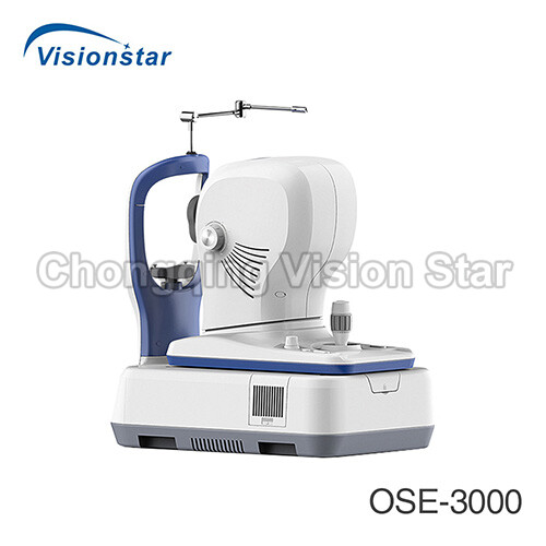
OSE-3000 OCT Optical Coherence Tomography
$0.00
OSE-3000 OCT Optical Coherence Tomography is equipped with Line Scanning Ophthalmoscopy, which helps take high quality HD images, and it is built with professional software, which will help the doctors to make analysis and get the report very easily.
OSE-3000 OCT Optical Coherence Tomography
Video:
Features:
MACULA
1. LSO:Equipped with LSO(Line Scanning Ophthalmoscopy)MOcean 3000 provides simultaneously high quality fundus imaging,which is easy for physicians to localize the lesion.
2. MACULA:Macula HD line:High definition OCT imaging reveals small lesions,OCT scan length can be switched between 6mm and 12mm.
3. Macula Six-line Radial: Having a glimpse of the retina with HD imaging and quick data analysis. Software Analysis: Retinal thickness analysis, Ganglion cell analysis, High definition OCT imaging with 5 images averaging.
4. Macula Cube: A point-by-point assessment of retinal thickness with a 500×100 dense cube
Software Analysis: Retinal thickness analysis,Progression analysis, 3D view,En-face analysis.
GLAUCOMA
1. For comprehensive glaucomaanalysis.MOcean 3000/3000 Plus offers two scan modes:glaucoma cube scan in macular area and glaucoma cube scan in disc area.Evenly distributed sampling rate with 200×200 A-scans provides reliable information for early glaucoma detection and management.
2. Glaucoma(Macular):Software Analysis:Ganglion cell analysis,Progression analysis.
3. Glaucoma(Disc):Software Analysis:RNFL analysis,Cup-disk analysis,Calculation circle and circle scan tomogram,Progressionanalysis,OU comparative analysis.
Informative Report: OU comparative analysis, Progression analysis report.
ANTERIOR SEGMENT
1. Anterior HD line:High-definition OCT imaging of the cornea enables localization of the Bowman’s layer, the interface between corneal stroma and and epithelium, Anterior Chamber Angle,Manual measurement is available.
2. Anterior Six-line Radial:The anterior segment scanning through 6 radial lines of equal length can be used to measure the central corneal thickness, Software Analysis, Corneal pachymetry,Manual measurement.
PERMIUM FUNCTIONS
1. En-face Analysis:En-face OCT provides ability to precisely localize lesions within specific subretinal layers.
Choroid View
IS/OS-Ellipsoid View
Mid-Retina View
VRI View
2. 3D En-face View
Network System: Remote Analysis System:Moptim provides a remote viewer software for displaying,enhancing, analyzing and saving digital images obtained with MOcaen 3000/3000plus.
Remote Diagnosis System: Customer scan are reviewed remotely by specialists at Big Picture Eye Health for over 45 eye pathologies.
The software immediately generates a customer report, including educational content and specialist referral if needed.
This optional module,which is developed and operated by Big picture Eye Health, can be connected to MOcean 3000 seamlessly.
Specifications:
| OCT Imaging | |
| Methodology | Spectral Domain OCT |
| Optical Source | Super Luminescent Diode(SLD), 840nm |
| Scan Speed | 36,000 A-Scans/s |
| Axial Resolution(Optical) | 5 Microns(optical), 2.7 Microns(digital) |
| Tranverse Resolution(Optical) | 15 Microns(optical), 3 Microns(digital) |
| A-Scan Depth | 2.3mm |
| Diopter Range | -20 to 20 Diopters |
| Scan Patterns | Macular: HD Line Scan(6mm or 12mm), 3D Scan(6mmx6mm), 6-Line Radial Scan. Disc: 3D Scan(6mmx6mm). Anterior: HD-Line Scan(6mm), 6-Line Radial Scan. |
| Fundus Imaging | |
| Methodology | Line Scanning Ophthalmoscopy(LSO) |
| Frame Rate | 10 FPS |
| Minimum Pupil Diameter | 3.0mm |
| Field of View | 47 Degrees |
| Software Analysis | |
| Macula | Retina Thickness Analysis; 3D View; En-face Analysis; Progression Analysis |
| Glaucoma | RNFL Analysis; Ganglion Cell Analysis; Cup-Disk Analysis; Progression Analysis; OU Comparative Analysis |
| Anterior Segment | Manual Measurement; Corneal Thickness Analysis |
| Others | DICOM Conformance; Remote Viewer Software Available |
| Electrical and Physical Specifications | |
| Weight | 29kg |
| Dimension | 450mm(L)*250mm(W)*450mm(H) |
| Input Voltage | AC 100V-240V |
| Frequency | 50Hz-60Hz |
| Rated Power | 90VA |
Chongqing vision star is a supplier of professional medical devices, including medical laboratory equipment, surgical equipment, ultrasound machines, radiology equipment, ophthalmology equipment, dental equipment, ENT equipment, obstetrics and gynecology equipment, neonatology equipment, rehabilitation room equipment, beauty equipment, hospital furniture, medical consumables and supplies. You can find a wide range of high quality devices that made in China for hospital or clinic. In the passed 20+ years, we have exported more than 150 countries, provided effective solutions for various types of medical system, and established long term cooperation with distributors and tenders in many countries. We sincerely support every customer with best products, prices, services, and warranty. For distributors and resellers, we can label your own logo on the devices if your order reaches MOQ. We can also help you to source more products that have not been listed on our website. Contact us to get more information.


