VXRSTAR Floor-mounted DR Machine
$0.00
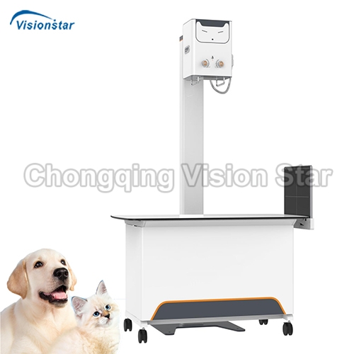
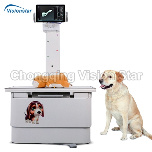
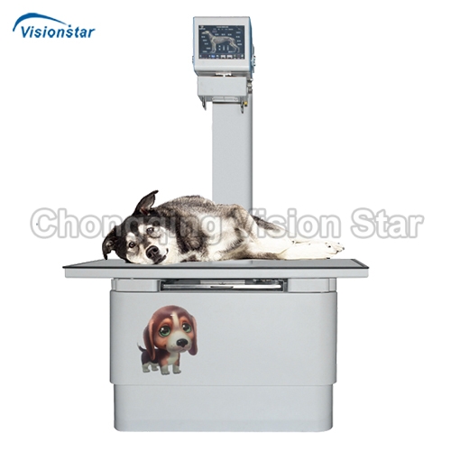
VXRSTAR Floor-mounted DR Machin
- Feature:
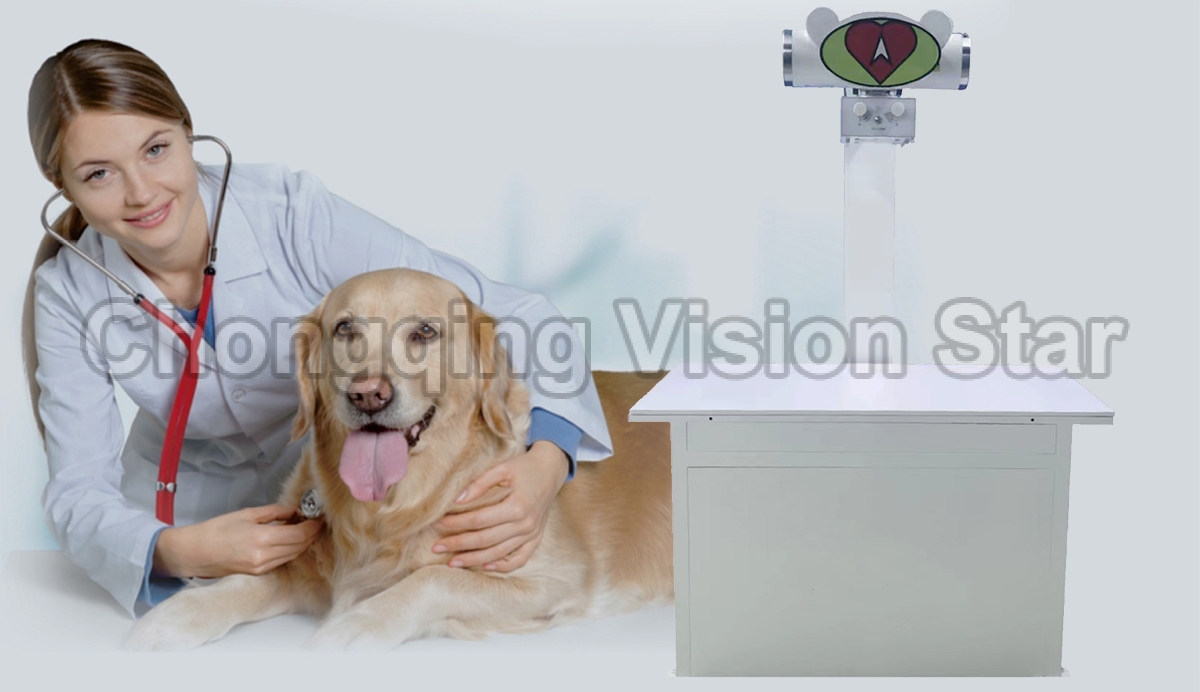
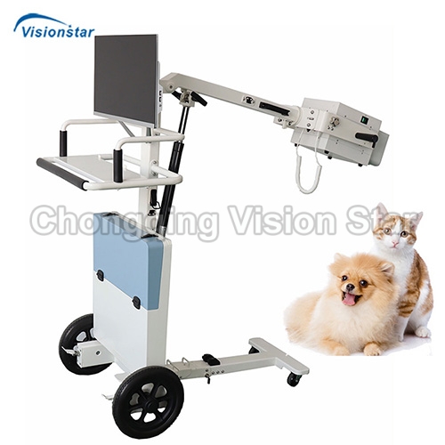
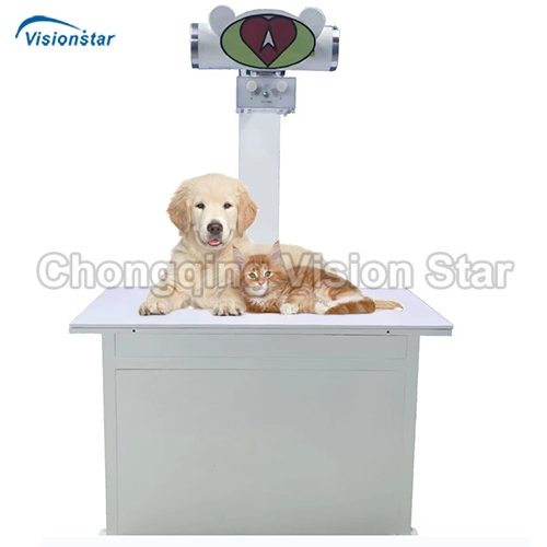
- * Pet DR is mainly used for full-body photography of pets, respiratory system, skeletal system, and various body positions, and can also be used for pet physical examination.
- * High performance
- * Excellent image
- * Support for advanced measurement tool
- * Low dose
- * Amorphous silicon cesium iodide flat panel detector
- * 17″ X 17″ Fixed Flat Panel Detector
- * Size 17″ x 17″, ultra-light design (<4.0kg), full field of view dynamic exposure sensing (F2AEDTM) technology, it is suitable for a variety of applications, such as the integration of X-ray photographic system requiring the use of detectors in different positions, or the upgrading of CR film system.
- * Advanced technology (high performance cesium iodide direct growth technology, optimized electronic digital signal processing technology) realizes the product’s high detection quantum efficiency, high spatial resolution, ultra-high dynamic range, and ultra-low noise level, ensuring high-quality images at low dose.
- * Configuration List
- * Set of cesium iodide amorphous silicon flat panel detector (17*17 inches large panel)
- * High voltage generator set
- * Set of camera bed and bulb column
- * A set of 4 foot brakes
- * A set of image acquisition software
- * Image acquisition workstation (computer host) a set
- * One workstation monitor (Philips 24 “monitor)
- * One hand switch
- * Set of beam limiter
- * Medical rotating anode X-ray tube assembly
- * High voltage cable set
- * One set of lead protective clothing
|
Technical Data |
|
|
Application |
For digital X-ray photography of animal head, cervical vertebra, limbs, chest, abdomen and other positions |
|
Main Unit |
|
|
Distance between focus and ground |
1500mm |
|
Distance between focus and receiver input screen (SID) |
1000mm |
|
Beam limiter requirements |
Photoelectric positioning, manual multi-blade field indicator with timing switch function |
|
Camera bed and machine |
1200m*600*1700mm(L*W*H) Universal wheel with brake function |
|
Detector |
|
|
Flat panel detector |
Cesium iodide amorphous silicon |
|
Pixel area (effective size) |
≥17inch x 17inch |
|
Pixel size |
≤140um2 |
|
Pixel matrix |
≥3K×3K |
|
Limit resolution |
≥3.6LP/mm |
|
A/D conversion |
≥16 bits |
|
Data output |
Gigabit Ethernet |
|
X-Ray Generator |
|
|
Generator |
High frequency high voltage technology |
|
Power supply |
Single-phase 220V± 10% |
|
Maximum tube voltage |
125KV |
|
High voltage operating frequency |
100kHz |
|
Camera tube voltage |
40 ~ 125kV, step by 1kV |
|
Loading time range |
1ms ~ 6300ms |
|
Maximum tube current |
200MA |
|
Maximum power |
16KW |
|
X-ray Tube |
|
|
Focal spot |
Small focus 1.0mm, large focus 2.0mm |
|
Power |
Maximum focus power 45KW |
|
Built-in temperature control switch, automatic protection |
|
|
Anode speed |
2700r/min |
|
Image processing Software |
|
|
The software has the image splicing function, supporting any number of images to splice, meeting the clinical needs of the overall observation of patients. |
|
|
Case management function: Perfect patient information registration function; Including patient information, examination information and image management; DICOM3.0 standard Worklist query service, one can query and download case data from PACS. |
|
|
Image acquisition: With detector calibration function, the detector can be calibrated directly from the software; Digital photography function, can configure positive and negative image acquisition; Real-time window width window position adjustment; Real-time edge enhancement; Select mirroring and rotation according to different position; Sharpness, contrast, noise reduction, frequency layer; Can display patient information, inspection information, equipment information, image information. |
|
|
Image processing: Window width, window position and Gamma adjustment, multi-point LUT curve adjustment; Positive and negative film conversion, image scaling, translation, mirroring, rotation, magnifying; Select the original display of the area, full screen display, histogram display, window width and window position adjustment; Image enhancement, noise reduction, different enhancement schemes can be set according to different parts, and the degree of enhancement can be adjusted; Image annotation function, including drawing lines, rectangular polygons, arrows and text; Prompt the number of images the system can store in real time; Supports multi-screen display of images. |
|
|
Image output: DICOM3.0 standard laser camera output, can easily choose a good solution (film size, layout) printing; DICOM sends SCU, which supports sending images to any following DI and backing up images to CD/DVD. COM3.0 PACS and workstations: Image backup capabilities. |
|
|
DR Image Acquisition Workstation |
|
|
Computer |
Memory: 8GB DDR4 Hard disk: 1TGB 3GSATA 7200 RPM hard disk Operating system: Windows 7 24 inch Philips LCD display |
|
Installation, Commissioning and Acceptance |
|
|
Responsible for free computer room design, line layout, equipment installation, commissioning, to ensure normal operation. Responsible for the free training of operators until they fully master the operation technology |
|
Chongqing vision star is a supplier of professional medical devices, including medical laboratory equipment, surgical equipment, ultrasound machines, radiology equipment, ophthalmology equipment, dental equipment, ENT equipment, obstetrics and gynecology equipment, neonatology equipment, rehabilitation room equipment, beauty equipment, hospital furniture, medical consumables and supplies. You can find a wide range of high quality devices that made in China for hospital or clinic. In the passed 20+ years, we have exported more than 150 countries, provided effective solutions for various types of medical system, and established long term cooperation with distributors and tenders in many countries. We sincerely support every customer with best products, prices, services, and warranty. For distributors and resellers, we can label your own logo on the devices if your order reaches MOQ. We can also help you to source more products that have not been listed on our website. Contact us to get more information.

