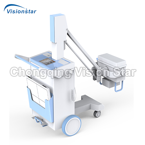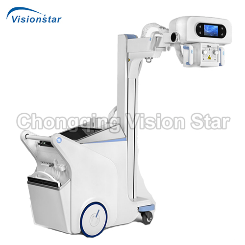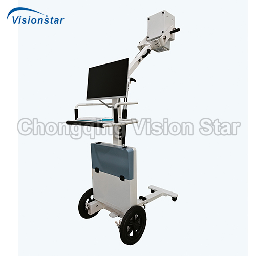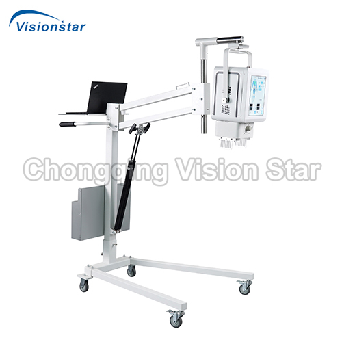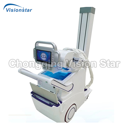
XMX32Y Mobile Digital Radiography System
$0.00
XMX32Y Mobile Digital Radiography System
Feature:
* Easy to operate, and flexible to move
* Automatic lifting columns can shrink the system to just more than four feet, allowing you to fully see the pathas you move the system anywhere.
* Powerful dual-motor drive allows you to move the system back and forth without difficulty.
* Large touch screen, Chinese handwritten input, interactive human-computer dialogue, bring a newinteractive experience and extraordinary work efficiency.
* Outstanding operation performance, flexible rotating expansion arm, broad imaging area, the tube canrotate around the column for a large range, shooting without dead corners.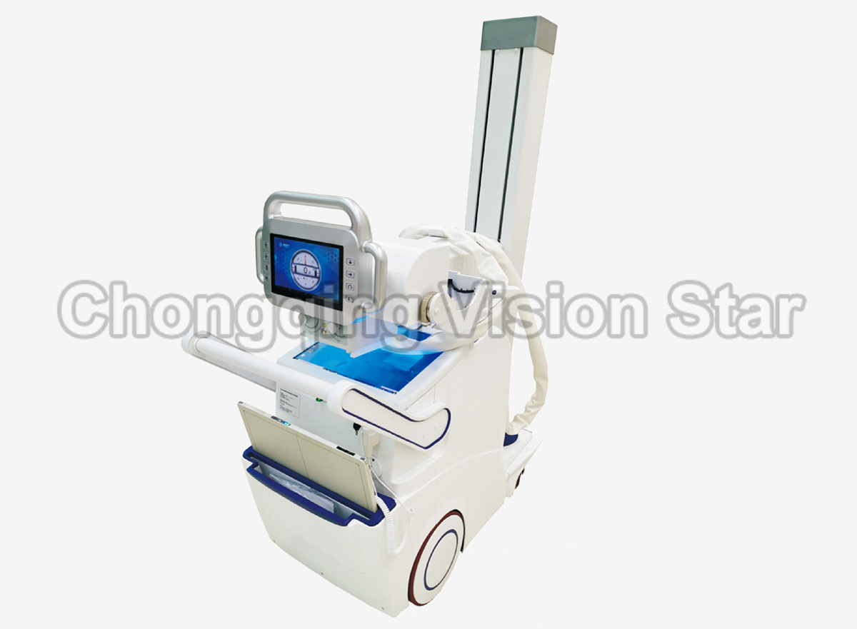
Work Station Functions for Image Acquisition, Processing and Diagnosis
(1) Standard DICOM3.0 image
(2) Work station functions for image acquisition: adjust or preset window width/ window level, local automatic window level, preset window width/ window level, positive and negative image flipping, image flipping, rotation, image magnification and roam, image interpolation edge enhancement, local magnification, restoration, image annotation, text annotation/number annotation, image marking, ruler line segment measurement, square and round measurement, arbitrary shape measurement, angle measurement, automatic electron shearing, image stitching, acquisition and display of exposure index.
(3) Software package for the special image acquisition control and software package for special parts protocol processing;
(4) Functions of patient management, image acquisition, image processing (image correction, image flipping, USM sharpening, image filtering), image observation (provide the tools for observation and measurement), image stitching;
(5) Image printing, DICOM printing, paper printing, manual printing for the displayed images, single button for marking and printing the images, optional for different printer, film format, and number of prints, print queue control, stop/start the preset;
(6) User personalization: showing the format and layout, default setting, toolbar setting, parts protocol enhancement filter;
(7) Image display: display configuration 1920×1080, HD display.
Work Station
(1) CPU: Intel i7,3GHz or above
(2) Host memory: ≥8GB DDR3 1600 high speed memory
(3) Hard disk: 1T/7200rpm large capacity and high speed hard disk
(4) Work station monitor: IPS LCD monitor
(5) Network card and network interface: 1000M network card, 1000M network interface, Jumbo Frames: 9K
(6) DICOM3.0 interface
(7) Windows 10 64 bit SP1 (Professional Edition or higher)
Configuration List
|
No. |
Parts Name |
Qty. |
Unit |
|
1 |
High voltage generator |
1 |
set |
|
2 |
X-ray tube assembly |
1 |
pc |
|
3 |
Flat panel detector |
1 |
suit |
|
4 |
Hanging stand |
1 |
pc |
|
5 |
Image work station |
1 |
suit |
|
6 |
Acquisition software |
1 |
suit |
|
7 |
Beam limiting stitching |
1 |
pc |
|
8 |
Grid |
1 |
pc |
|
Technical Data |
XMX32Y |
XM50Y |
|
Flat Panel Detector |
||
|
Type of the detector |
TFT monolithic amorphous silicon |
|
|
Panel size of the detector |
430mm*430mm |
|
|
Efficient imaging size of the detector |
17*17 inch |
|
|
Acquisition pixel matrix of the detector |
≥3072×3072 |
|
|
Pixel pitch of the detector |
≤146 um |
|
|
Spatial resolution of the detector |
3.4LP/MM |
|
|
Pixel gray scale |
16 Bit |
17 Bit |
|
Time of image acquisition and transmission |
3s |
|
|
High Frequency and High Voltage Generator |
||
|
Max. Output power |
30kw |
50kw |
|
Input power supply |
220 VAC single phase |
|
|
Output voltage |
40~150kV |
|
|
Tube current range |
10~320mA |
10~630mA |
|
Exposure time range |
1ms-10000ms |
|
|
mAs range |
0.1mAs~1000mAs |
|
|
Support AEC、APR automatic exposure |
||
|
Diagnostic self-test and display |
||
|
X-ray Bulb Tube Assembly |
||
|
Focus |
0.6mm/1.5mm |
|
|
Nominal electric power |
21kw/46kw (50Hz) |
|
|
Max. KV |
150kV |
|
|
Anode target angle |
12° |
|
|
Column |
||
|
Rotation angle range of the column |
145°~180°, deviation±2° |
|
|
Up and down rotation angle of the bulb tube and the beam limiting device on horizontal axis |
-90°~45°, deviation±2° |
|
|
Vertical moving stroke of the X-ray tube assembly focus |
95cm, deviation ±2cm |
|
|
Rotation angle in horizontal direction of the bulb tube and the beam limiting device |
greater than ±180° |
|
|
The Grid and the Beam Limiting Device |
||
|
The grid material |
Aluminum based grid |
|
|
Size |
18×18 inch (48×48cm) |
|
|
Grid ratio |
10:1 |
|
|
Wire/inch |
215C |
|
|
Grid Focal length |
1000mm |
|
|
Light field, multi-blade DR beam limiting device |
||
Chongqing vision star is a supplier of professional medical devices, including medical laboratory equipment, surgical equipment, ultrasound machines, radiology equipment, ophthalmology equipment, dental equipment, ENT equipment, obstetrics and gynecology equipment, neonatology equipment, rehabilitation room equipment, beauty equipment, hospital furniture, medical consumables and supplies. You can find a wide range of high quality devices that made in China for hospital or clinic. In the passed 20+ years, we have exported more than 150 countries, provided effective solutions for various types of medical system, and established long term cooperation with distributors and tenders in many countries. We sincerely support every customer with best products, prices, services, and warranty. For distributors and resellers, we can label your own logo on the devices if your order reaches MOQ. We can also help you to source more products that have not been listed on our website. Contact us to get more information.


